Ultrasound scanning, carried out in each trimester of pregnancy, allows us to assess the condition of the fetus and identify possible defects. One woman out of two dozen prefers to refuse this procedure. The expectant mother relies on the mistaken belief that ultrasound can harm the baby. In fact, everything is exactly the opposite. If your doctor prescribes an ultrasound scan at 18 weeks or at any other time, you must follow these recommendations.
-
What happens to a baby at 18 weeks?
It's the fifth month of pregnancy. The belly of the expectant mother is growing by leaps and bounds. It is already quite difficult to hide it from strangers, but keeping a secret is still possible if you really want to. The uterus resembles a medium melon. It rises several centimeters above the womb and has already reached the navel.
At eighteen weeks, many expectant mothers already feel the baby moving. If this is not the first pregnancy, then noticeable tremors can be felt two weeks earlier. Slender women talk about soft, unobtrusive shocks, reminiscent of bursting bubbles. Expectant mothers “in the body” may not yet feel them, but there is nothing wrong with that. Be patient, after 2-4 weeks you will feel the baby's movements.
At 18 weeks of gestation, the fetus reaches the size of a ripe mango. The baby's limbs are fully formed, now they just have to grow. The fingers have phalanges that have a unique pattern. The eyes of the unborn child still remain closed, but they clearly distinguish the light. If you direct a bright beam at the belly, the baby will try to cover its face with its hands. The brain continues to increase in volume. This stage is very important for a small organism. The baby needs constant contact: talk to him, stroke his tummy. Be sure to involve the future dad in this.
Ultrasound in the fifth month of pregnancy
Ultrasound scanning in expectant mothers can be performed in two ways: transvaginally and transabdominally. Depending on individual characteristics and the indications for the study, the doctor chooses the most appropriate method.
Transabdominal scanning is performed with a special sensor using conductive gel. During the manipulation, the woman does not feel discomfort except for the apparatus moving across the abdomen. The procedure lasts from 5 to 30 minutes. A transabdominal study is performed to study the fetus: determine its size, obtain information about development and behavior.
Transvaginal ultrasound is needed when the first method cannot provide the required amount of information. For example, if the embryo is rotated in a certain way and does not allow the specialist to measure the necessary organs and parts. Also, a transvaginal sensor can give more information about the condition of the cervix than its counterpart.
If the expectant mother has cervical insufficiency or premature opening of the internal os is suspected, then an examination is performed through the vagina.
Why is ultrasound prescribed: indications
During the entire gestation period, a woman will undergo three main scans. These are called screening studies. The results obtained after the first such diagnosis are considered very important and the most revealing. If these data do not fit into established and generally accepted norms, then the doctor may prescribe a routine ultrasound at 18 weeks of pregnancy. During this period, it is much easier to assess the child’s condition.
Why don’t they want to wait the required 20 weeks for the second screening? The fact is that if it is necessary to terminate a pregnancy, it is much easier to carry out such manipulation at a period of 18-19 weeks. The procedure will entail less negative consequences than an abortion at 22 weeks. So, the main indication for performing a routine ultrasound at 18 weeks is bad results first screening.

Ultrasound examination may have other reasons for carrying out. Scans are sometimes performed for emergency reasons. The doctor may prescribe a diagnosis for you if you have the following complaints:
- bleeding from the reproductive organ or beige-brown spotting;
- pain in the lower abdomen;
- discrepancy between the volume and size of the abdomen and the expected period;
- abnormalities in the location of the placenta, previously identified.
In addition to ultrasound, the woman is prescribed additional tests that can tell about the reasons for this or that deviation.
What can you see during a scan?
An ultrasound at 18 weeks of pregnancy is designed to evaluate the development of the embryo. The specialist takes measurements of body parts: arms and legs, head circumference and tummy. Attention is drawn to the presence of internal vital organs. At this stage, the nasal bone and the thickness of the collar space are no longer indicative, so attention is not focused on them.
During ultrasound manipulations, you can see the condition of the uterus. The doctor determines the location of the placenta and determines its maturity. The umbilical cord must be examined (the number of vessels and its length are studied). It is important to establish the condition of the internal pharynx. In the fifth month of pregnancy it should be tightly closed.
During an ultrasound performed at week 18, developmental defects that were not previously noticed can be detected. If the first screening showed poor results, then the next examination can already give a clear assessment of the developing baby.
Gender
Many couples wonder: will an ultrasound at 18 weeks of pregnancy show the gender of the baby? The answer to this will most likely be positive. An ultrasound scan performed by a competent specialist using a modern device can determine the gender of a child with 100% certainty.
Some women preparing for motherhood specifically go for research to find out the gender of the baby. You shouldn't do that. Increased interest will be satisfied, but frequent scanning is not the best for a normally developing pregnancy.
There are also situations in which it is necessary to find out the sex of the baby in advance. This is necessary for inherited autoimmune diseases or other pathologies.
What do the genitals of an embryo look like at this stage? In girls, the labia can be clearly seen. But from a certain angle they can resemble testicles. Therefore, some experts may predict that you will have a boy, but the birth will be a girl. It is much more difficult to confuse the male and female genders. At this stage, the male embryo has a clearly visible penis.
Note: the testicles of a male fetus at 18 weeks of pregnancy have not yet descended into the scrotum. This will happen closer to childbirth. If the doctor shows you the testicles and promises the birth of a boy, be wary: most likely, the result is incorrect.
What do the resulting numbers mean?
 After monitoring, the ultrasound diagnostic specialist will issue you a protocol. The form indicates all the values and characteristics of the fetus obtained during the ultrasound. It also sets out norms that are considered generally accepted.
If your values fall within the standard range, then we can talk about a normal pregnancy. Any deviations in one direction or another must be recorded by a gynecologist. The doctor monitoring your pregnancy can make a specific diagnosis based on the findings.
You should not try to decrypt it yourself. Most likely, you won't succeed. If you find values that do not fit into the established framework, you will only worry once again, which is extremely undesirable. At 18 weeks of pregnancy standard values will be as follows:
- the weight of the embryo is about 200 grams, and its length is from 12 to 15 centimeters;
- the fronto-occipital zone is from 4.9 to 5.9 cm, and the BPR (biparietal size) is from 3.7 to 4.7 cm;
- the circumference of the future baby's tummy is 10.4-14.4 cm, and the circumference of the head is 13.1-16.1 cm;
- the length of the femur is about 3 cm, and the length of the tibia is 2.5 cm;
- the forearm is approximately 2 cm, and the humerus is half a centimeter larger;
- the degree of maturity of the placenta is zero, the thickness at this stage has not yet been measured;
- the volume of amniotic fluid ranges from 80 to 220.
Depending on the equipment, normal ultrasound readings may vary slightly.
Possible deviations
If the diagnostics revealed indicators that do not fit into the norms, then you should not immediately panic. Deviations can be natural and pathological. You cannot make a diagnosis based on ultrasound alone. To confirm or refute suspicions, additional studies are carried out: dynamic scanning; Dopplerography; blood test for hormones, antibodies, and so on.
If the size of the embryo does not fit into the established standards, then this may be hereditary. Parents of short stature and thin physique do not give birth to heroes. The baby may be larger than the stated norms. This often happens in obese couples or women with diabetes.

Polyhydramnios and oligohydramnios are also deviations. In the first case, the cause may be chronic diseases of the mother or Rh conflict. If you had an infection during pregnancy, this is also accompanied by polyhydramnios. Oligohydramnios is a more dangerous phenomenon. It becomes a consequence of fetal defects, for example, lack of kidneys. Doctors still cannot reliably establish the causes of this pathology.
Deviations from the norm may occur due to congenital anomalies. They are often accompanied by a delay intrauterine development, growth retardation, non-compliance with normal values. If the fetus is missing any vital organ, then this is an indication for termination of pregnancy. In other cases, possible drug correction or surgical intervention is carried out.
Let's sum it up
If your pregnancy is proceeding smoothly and does not have any abnormalities, and the results of the first screening are good, then you will not be scheduled for an ultrasound at 18 weeks. You should not go for diagnostics on your own just to find out the sex of the baby. Wait for the second screening, which falls between 20 and 24 weeks.
Coming for an ultrasound, each time the expectant mother strives to get a better look at her baby and get to know him. Ultrasound at 17-18 weeks of pregnancy is especially interesting. The seventeenth week is considered the beginning of the fifth month of fetal development. This is a time of relative calm, the absence of any negative feelings. In some cases, a pregnant woman may experience negative changes such as:
- increased sweating;
- bleeding gums;
- the appearance of copious vaginal discharge.
During this period, doctors recommend that the expectant mother get plenty of rest and try not to bother herself with various worries. A woman may feel some pain due to the fact that the uterus grows upward. She begins to worry about heartburn, frequent urination, and shortness of breath.
Since eighteen weeks, a pregnant woman has been bothered by pressure from the inside on the navel. This is due to the rapidly growing uterus. By this time it has already reached the size of a melon. At 17–18 weeks, the expectant mother’s belly is already clearly visible.

Starting from the 17th week, the baby’s immune system is already activated, which is manifested in the fetus’s production of its hormones (immunoglobulin, interferon). Under his skin, fat is formed, which he will need to regulate heat exchange in the body. By this time, the baby’s heart is almost formed and performs its important function.
From the 17th week, the formation of permanent fetal teeth begins. In girls, the uterus is forming. The adrenal glands produce hormones, and the pituitary gland is active. The baby can already hear and is able to distinguish between his mother’s emotions. This development is characteristic of the fetus without the presence of any pathologies. If a child. the mother has no special indications for ultrasound diagnostics, then at this stage (17 weeks) ultrasound is not prescribed.

At the 17th week, the baby’s movements are still barely perceptible, which cannot be said about the 18th week. At this time, the baby rattles quite noticeably, often and strongly. The norm of movements by this time is 4 – 8 per hour. But each organism is individual, so there may be various deviations from the norm.
By the 18th week, the baby’s arms and legs are already fully formed, and the phalanges are already visible on the fingers. The child already has his personal fingerprints, their unique pattern. During this period, the development of the brain occurs, a reaction to light and sounds is noted.
Developmental norms in the fetus at 17, 18 weeks

A baby at 17-18 weeks is already quite big. The normal height at 17 weeks is 12 cm, the baby’s weight reaches 100 grams. An ultrasound at 17–18 weeks shows the development of the fetus and the presence/absence of pathologies.
Normal fetometry indicators at week 17 are:
- BPR. Biparietal size is approximately 34 – 42 mm;
- OG. Head circumference is 112 – 136 mm;
- LZ. The fronto-occipital size is 41 – 49 mm;
- coolant. Abdominal circumference ranges from 121 to 149 mm.
As for the size of the long bones of the fetus, they are normally:
- forearm bones – 15 – 18 mm;
- femurs – 20 – 28 mm;
- shin bones – 15 – 21 mm;
- humerus – 15 – 21 mm.
In addition to the size of the fetus at 17 weeks, the specialist also studies ultrasound examination condition of the placenta and the child’s brain.

By the 18th week, qualitative changes occur in the baby’s body. By the end of the month, his weight will reach 150 grams, and his height will be about 12.5 - 14 cm.
- BPR. Biparietal size is approximately 37 – 47 mm;
- OG. Head circumference is 131 – 161 mm;
- LZ. The fronto-occipital size is 49 – 59 mm;
- coolant. Abdominal circumference ranges from 104 to 144 mm.
As for the size of the long bones of the fetus, they are normally:
- forearm bones – 17 – 23 mm;
- femurs – 23 – 31 mm;
- shin bones – 23 – 31 mm;
- humerus – 15 – 21 mm.

From the 18th week, a specialist can already determine the gender of the unborn baby and show it to you in a photo. At 17–18 weeks of pregnancy, the specialist not only examines the external and internal organs of the child, he also receives information about:
- number of fruits;
- condition, location of the placenta (it should be localized on the back wall of the uterus);
- the cervix (its length should not be shorter than 30 mm, the external and internal pharynx should be closed);
- amount of amniotic fluid;
- condition of the uterus (the specialist must make sure that there is no hypertonicity of the uterus.
- presentation of the fetus (normally it should be cephalic at this stage).
Preparing for diagnostics

At 17–18 weeks of pregnancy, diagnosis can be carried out in two ways:
- transabdominal. The sensor is moved over the surface of the abdomen after preliminary lubrication with a special gel;
- transvaginally. With the insertion of a sensor into the vagina.
Preparation for an ultrasound examination is practically not needed if it is performed transvaginally (except for individual hygiene of the genital organs). If the diagnosis is performed transabdominally, then the pregnant woman is recommended to:
- do not eat heavy food on the day of the examination;
- do not consume foods that cause increased gas formation;
- Fill your bladder before testing.
![]()
If an ultrasound is prescribed at 18 weeks, there is no need to fill the bladder; the volume of amniotic fluid is sufficient for a detailed study of the structure and size of the unborn baby. In addition to conventional ultrasound examination, a specialist may prescribe Doppler testing. This method diagnostics allows the doctor to study in detail the blood flow inside the uterus and fetal vessels. There is no special preparation for this examination.
The price of an ultrasound examination performed transvaginally is approximately 1000 - 1500 rubles.
![]()
It is recommended to carry out the study in the second and third trimester, but if there are special indications, this diagnosis can be carried out even later. early stages. You should come for the examination with an empty bladder. It is also necessary to avoid bloating.
The cost of a transabdominal ultrasound is 900 – 1100 rubles.
You need to take a diaper with you to the examination. If you need anything else (gel, wipes), the doctor will warn you in advance. The cost of diagnostics may vary depending on the type of medical institution, the device used, and the qualifications of the specialist.
Is it harmful to have an ultrasound scan at 17–18 weeks of pregnancy?

At 17 weeks, a planned study is usually not prescribed. According to the plan, the diagnosis takes place at 18 weeks. A specialist may refer a pregnant woman at 17 weeks for an ultrasound examination in the following cases:
- Failed diagnosis at 12 weeks (first trimester).
- The expectant mother has serious chronic diseases.
- Exposure of pregnant women to acute infectious diseases.
- Unsatisfactory biochemical screening of the first/second trimester (deviation from the norm of indicators such as: PAPP, free estriol, beta-hCG, alphafetoprotein).
- The requirement of a married couple to carry out diagnostics to exclude chromosomal abnormalities and fetal malformations.
- Weakness of the cervix.

No harmful effects of ultrasound on fetal development were found. There is no need to worry again if you are prescribed an additional ultrasound examination at 17 weeks. This diagnosis will not affect your baby’s condition, but it will provide the specialist with comprehensive information about existing disorders. At this time, the following pathologies may be detected in the child:
- abruption, placenta previa;
- developmental delay;
- structural anomalies;
- fetoplacental insufficiency;
- a lot/little water;
- premature aging of the placenta;
- failure of the scar on the uterus, which remained after a previous operation (cesarean section).
Timely identification of such pathologies can be decisive in maintaining pregnancy. Be sure to go through all the tests that your gynecologist directs you to do. Thanks to a thorough examination of the condition of the fetus and the mother’s reproductive organs, a specialist will help solve all problems that arise during pregnancy as soon as possible.
In the fifth month of pregnancy, a woman gains significant weight - about 5-7 kilograms. Since the bulk of the weight gained comes from amniotic fluid, the placenta and the baby itself, there is no reason to worry. An ultrasound at 18 weeks of pregnancy will help identify possible deviations in the development of the fetus, and, with a successful combination of circumstances, find out the gender of the unborn child.
The ultrasound examination procedure performed by specialists is absolutely safe and does not harm the fetus or mother. A relatively mature baby is already resistant to various influences, and ultrasonic vibrations cannot harm him.
Why do an ultrasound at this stage?
Parents are often ready to undergo the procedure themselves - this will help to find out the sex of the child. However, this is only possible under certain conditions: the baby must be in a position in which it is possible to see his genitals (scrotum or labia). The formation of the genital organs occurs in the period from 17 to 20 weeks, and the probability of determining the sex of the child during examination during this period is quite high.
There is an opinion that the fetus perceives noise from ultrasonic vibrations in the same way as a person perceives a loud sound. This can cause the baby to become restless, which can negatively affect his well-being. But as numerous practice has shown, such a judgment has no confirmed justification, and an ultrasound scan at 18 weeks can be done without worrying about the baby’s health.
The main goal of research at this time is to check the condition of the cardiovascular system, heart and determine the size of the frontal and occipital parts of the head. An ultrasound will reveal possible abnormalities in the development of the child’s heart and blood vessels, as well as assess the level of its development.
At eighteen weeks, the weight of the fetus can be up to 250 grams, and its growth rate is 14-15 cm. During this period, the development of the fetus occurs without significant changes - all the organs and tissues of the baby are in the process of growth.
Also, ultrasound readings can promptly identify pathologies that provoke cases of miscarriage or stillbirth of children, abnormal development of the placenta (its premature aging, abruption, etc.). If the ultrasound reveals even a slight deviation from the accepted norm, then measures will be taken to maintain the pregnancy.
How does the baby feel at this stage?
An ultrasound at 18 weeks can show the baby's first smile and his attempts to know himself and the world around us. You can often see on the monitor how the baby sucks his thumb or yawns. The uterus, which has grown to the size of a melon, gives the child a place to “travel” - he can already push off from its walls with his feet and periodically disturb the mother with his activity.
In the fifth month, the fetus begins to form the cerebral cortex and its individual areas responsible for auditory perception, vision, taste buds and touch. He begins to hear his mother’s voice and feel her condition, reacting in his own way to her anxiety.
During this period, the child is characterized by:
- first conscious movements;
- facial expressions;
- actions that are a reaction to the mother's anxiety;
- active movements when the mother eats tasteless food.
The activity of the fetus manifested at this stage is the norm. Many mothers begin to talk to the child - by the fifth month he can already hear her voice.
How does a woman feel at 5 months pregnant?
The rapid growth of the abdomen leads to a change in the gait of the expectant mother: when walking, she begins to lean forward, which is typical for all pregnant women. To relieve the strain on your lower back, it is recommended to wear special underwear designed for pregnant women.

At 18 weeks of pregnancy, women may experience symptoms of forgetfulness; it often becomes difficult for a woman to concentrate on anything for a long time. There is no need to be upset - for most pregnant women this is the norm. With the birth of the child, symptoms usually disappear and can only return during the next pregnancy. Experts recommend taking a number of vitamins that help improve brain activity. At this stage, women begin to complain of the first manifestations of back pain and pulling sensations in the lower abdomen.
An ultrasound performed at week 18 is necessary to assess the child’s growth, development, and sex determination. The specialist checks the received data for compliance with the development norms that are established for a certain period. The sonologist will tell the parents the sex of the baby if he can clearly examine the already formed genital organs of the fetus. Sometimes the gender remains a mystery until the next ultrasound. But the main purpose of ultrasound during this period is to diagnose defects in the development of the cardiovascular system and heart.
By 18 weeks, the fetus has grown to 14–15 cm, by which time the baby’s weight is already about 180 grams. There are no significant changes in the child's development during this period. The skeleton, muscles, and all internal organs are in a state of growth. They continue to get stronger.
Almost all expectant mothers feel fetal movements by the 18th week of pregnancy. Active movements of the baby appear in the evening, when the mother eats a tasteless product, as well as when the woman experiences negative emotions.
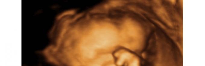
During an ultrasound, you can see how, at 18 weeks of pregnancy, the baby learns about the world around him. It can push itself from wall to wall in the uterus. The fetus feels its face, sucks thumb. All these moments are clearly visible during a 3D ultrasound; if desired, they can be captured and kept as a keepsake.
Expectant mother at 18 weeks of pregnancy
The expectant mother is usually overwhelmed with positive emotions at 18 weeks of pregnancy. The woman already clearly feels the movements and beatings of the child. A bunch of tests are left behind. The main thing is that by the 18th week, parents already know the gender of their baby. In most cases, it is possible to find out the gender, the deadline already allows.
Sometimes the expectant mother may become nervous, especially if the fetus was moving and then suddenly stopped. In this situation, you need to contact an obstetrician-gynecologist. He will be able to listen to the fetal heartbeat and also check the likelihood of abnormal development through ultrasound.
By the 18th week of pregnancy, the uterus reaches the size of a melon. Naturally, the tummy increases noticeably. During this period of pregnancy, walking outside is very beneficial. Fresh air necessary for the fetus to thrive, as well as for the mother.
The expectant mother can gain 5–7 kg by the 18th week of pregnancy. There is no need to worry too much about this because most of the kilograms fall on the fetus, placenta, and amniotic fluid. At this stage of pregnancy, specialists check the amount amniotic fluid. Its excess or deficiency is considered a deviation from the norm.
Due to the rapid growth of the abdomen, the pregnant woman leans slightly forward when walking. This gait is characteristic of all women carrying a child under their hearts. To ease the increased load on the back, it is recommended to wear special panties and shorts.
Forgetfulness and memory impairment often accompany pregnancy; you should not worry about this, for many this is the norm. But after birth, these symptoms should disappear. To improve memory, experts recommend taking special vitamins.

Baby development
At 18 weeks of pregnancy, the baby has about 100,000 cells in its brain. It is these cells that are necessary for connecting the brain with nervous system body. It is for the normal development and formation of all systems during pregnancy that it is necessary to take all recommended vitamins and their complexes. The baby needs them in order for the birth and development of all vital functions necessary for the body to occur.
;
Meconium continues to form inside the fetus. This substance is original feces. It is formed from those remnants of amniotic fluid that have not been digested. The baby will pass meconium for the first time after birth (the next day). If a bowel movement occurs inside the mother’s body, this indicates fetal asphyxia.
To check amniotic fluid, it is necessary to carry out a procedure such as amnioscopy. The woman sits on the gynecological chair, and the specialist examines the cervix through the amniotic sac and also evaluates amniotic fluid.
At 18 weeks of pregnancy, a second trimester ultrasound is performed. During this period, the baby's genitals can be seen very clearly. Parents can find out the gender of the child if he does not hide his intimate place. Sometimes gender determination is postponed to the next ultrasound. From the 18th week, the child’s body begins to produce myelin (the protective covering of the nerves).
All the sensory centers necessary for life are formed in the fetal brain:
- Hearing;
- Vision;
- Smell;
- Taste;
- Smell.
By the 18th week of intrauterine development, all organs necessary for life have already been formed. During this period of pregnancy, the fetus experiences their further development. Normally, by the 18th week the following features are characteristic:
- The fingerprints are already formed;
- Strengthening of the skeletal system is observed;
- The rudiments of milk and molar teeth are fully formed;
- Adipose tissue is rapidly formed;
- Formation of folds in the skin;
- The baby's skin is protected with a special lubricant. It protects against prolonged exposure to water;
- The retina of the eye develops. The eyes are closed, but the baby is able to distinguish external lighting;
- Centers for hearing, vision, smell, and taste are formed in the brain;
- The first feces are formed in the intestines, which will be excreted from the body after birth;
- The genital organs of the fetus are formed. Its gender can be determined with 99% accuracy.
Norms of fetal development by the 18th week of pregnancy
During this period, ultrasound measures such important indicators of the child’s development. Normally they are:
- Head circumference. Normal exhaust gas is 130 – 160 mm;
- Biparietal size. BPR – 36 – 46 mm;
- Abdominal circumference. Coolant – 102 – 114 mm;
- Fronto-occipital size. LZ – 48 – 58 mm.
During this period, the length of the bones must be determined by ultrasound. The following indicators, which are given taking into account the norm, are also of great importance for specialists:
- Femoral – 22 – 32 mm;
- Shin – 22 -32 mm;
- Shoulder – 15 – 20 mm;
- Forearms – 17 – 24 mm.
These indicators are indicated for normal fetal development, but if there are any significant deviations from the norm, it is worth conducting an additional ultrasound and testing.
Preparing for an ultrasound examination
To undergo diagnostics at the 18th week of pregnancy to the expectant mother no special training is needed anymore. There is no need to fill your bladder. The specialist will clearly examine the fetus with an ultrasound through the amniotic fluid, which is already quite sufficient. An empty or full stomach will not affect the diagnostic results.
In addition to the usual ultrasound, from the 18th week of pregnancy, doctors prescribe Doppler ultrasound. It is necessary to assess blood flow in the following organs:
- Uterus;
- Placenta;
- Vessels of the fetus.
Ultrasound of this period will also provide information about:
- Number of fruits;
- Location of the placenta;
- Presentation of the baby.
Prenatal screening is also carried out during this period. This study is necessary for those women who have been identified as likely to develop chromosomal, genetic abnormalities of the fetus. Most often these are pregnant women aged 17, 35 years, or women who had genetic diseases in their family. In such cases, gender determination is considered necessary.